Mapping the Mind: A Neuroscientist's Expedition into the Unknown

When neuroscientist Ed Boyden first approached the National Institutes of Health for funds to develop expansion microscopy, he was rejected. So he applied again. Second time around he was also rejected, but undeterred, he applied again and again and again and again.
"Many NIH grant proposals were rejected one after the other as people just didn't believe this could work," says Boyden, from the MIT Media Lab and the McGovern Institute for Brain Research. "But I always knew expansion microscopy was going to be really useful."
"We've now taught hundreds of groups how to use it, and I really do think it's going to be applied across all of biology because you can use it to see very beautifully the building blocks of life," he adds.
Expansion microscopy is like nothing else. Conceived during a research group brainstorming session, the method solves the problem of not being able to distinguish densely packed features in cells by making the tissue bigger, rather than improving microscope optics. The end result is that nanoscale structures can be scrutinized, even when using your everyday confocal microscope.
To swell tissue, sodium acrylate is infused into chemically fixed tissue, with polymerization agents then added to form a polymer network within the sample. The tissue-polymer sample is then chemically softened to homogenize mechanical properties, and finally rinsed in water to trigger linear expansion. Then, fluorescent labels are attached to specific targets, such as neurotransmitters, receptors, and ion channels, ready for imaging.

After expanding the fruit fly brain to four times its usual size, lattice light-sheet microscopy is used to image all of the dopaminergic neurons (green).
Already, researchers worldwide have been using the method to expand tissues from rats, mice, zebrafish, fruit flies, even bacteria, from four to ten times, reaching up to 30-nm resolution. Boyden and colleagues have also optimized expansion microscopy for pathology to enable accurate computational discrimination between high- and low-risk lesions on cancer biopsies. And at the same time, they have reached twenty-fold expansion and 25-nm resolution by expanding mouse brain slices twice.
While these resolutions match those of superresolution imaging methods such as stimulated emission depletion (STED) microscopy, the story of expansion microscopy hardly stops here. Boyden is adamant there is no fundamental limit to his method, which he believes can be used to expand biological samples over and over again.
"What if one day we could map all of the biomolecules throughout an entire cell?" he asks. "That would mean getting resolution greater than any previous optical microscopy method and expanding the cell not just 100 or 1000 times in volume, but a million times in volume, or more."
Genuinely feasible? Boyden says yes. Anytime soon? Absolutely.
"This is just a matter of chemistry and we think we can improve this until we are able to expand individual biomolecules away from each other," says Boyden. "So this is one of our goals: to get down to single-molecule precision in the next couple of years."
Given the swift successes of expansion microscopy and his determination to understand the brain at a deeper level, Boyden took his technology to Nobel Laureate, Eric Betzig, from the Howard Hughes Medical Institute's Janelia Research Campus, earlier this year.
By combining expansion microscopy with Betzig's incredibly fast high-resolution lattice lightsheet microscope, the pair imaged an entire fruit fly brain in a blisteringly fast 62.5 hours, showing some 40 million synapses in detail down to 60 nm.
This level of detail doesn't yet match that obtained with an electron microscope, but efforts to fully map the neurons and synapses of the fly brain with electron microscopy have taken decades with the efforts of dozens of researchers. Boyden reckons his method can be honed to scan an entire fly brain in just 12 minutes.
In addition to the fly brain, the researchers have also imaged large-scale circuits across a mouse cortex. At the time, Betzig said he "couldn't believe the quality of data" he was seeing.
"The imaging speeds we achieved are about one thousand times faster than the nearest best superresolution technology, and we think it's just a matter of, say, using more cameras and optics to go ten thousand times faster, and even more," says Boyden. "We're so excited about making scalable brain mapping a reality that this is now one of my group's main focuses."
The implications of swiftly imaging larger and larger volumes of the brain in 3D and with single-molecule precision are almost beyond imagination. If an incredibly detailed map of an entire brain can be made, this would provide a crucial step towards developing a brain simulation.
And since deeply understanding the brain has always been at the forefront of Boyden's mind, he has already developed additional technologies necessary to get him there. Long before expansion microscopy reached laboratories around the world, his first breakthrough brain analysis method, optogenetics, had already rocked the world of neuroscience.
Boyden conceived the idea of optogenetics with Karl Deisseroth, while each was at Stanford. The young students wanted to control neural circuits and decided that, thanks to its speed, light was the best means to deliver energy into the brain to then switch neurons on and off.
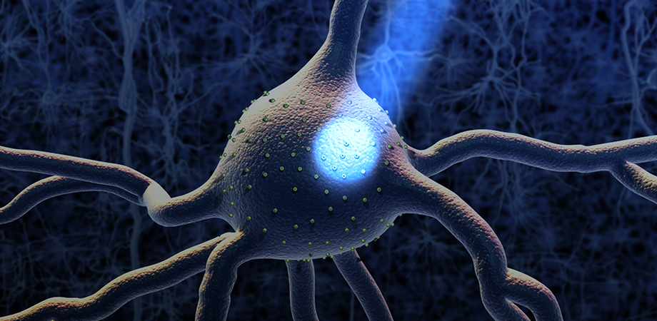
They went on to use blue light to control neurons in living brain tissue that had been genetically modified to express light-sensitive ion channels. The light modulates the membrane potential of the cells, switching transient electrical signals, which are the basis of neuronal computation, on and off.
Fast forward a decade-and-a-half, and thousands of research groups are using optogenetics while start-ups are pursuing optogenetics research in humans. For example, the method has been used to restore vision in mice, suppress Alzheimer's-related beta-amyloid plaque production in mice brains, and even trigger aggressive behavior in mice.

Top left: A forest of dendritic spines protruding from the branches of neurons in the cortex (Gao et al., Science 2019). Top right: A subset of pyramidal neurons (orange) in the mouse primary somatosensory cortex. The dendritic spines associated with the postsynaptic protein Homer1 are highlighted in yellow (MIT). Bottom left: Organelles of various shapes and sizes (colored areas) inside mouse neurons imaged by merging expansion microscopy with lattice lightsheet microscopy (Gao et al., Science 2019). Bottom right: Dopaminergic neurons in the ellipsoid body of a fruit fly brain, color-coded by 3D depth (MIT).
Meanwhile, Boyden has also developed fast-switching molecules that can be activated with red light for lower light scattering and minimal phototoxicity. Importantly, these molecules can be used to control neurons deeper within tissue.
"With our colleague Valentina Emiliani, we've reached single-cell submillisecond precision, so we're getting to the fundamental spatial and temporal limits of optogenetics," points out Boyden, "We will continue to refine the method, but it is nearing the endgame."
The rate of optogenetics development has been breathtaking, but in addition to controlling the neurons within a live brain, Boyden wants to watch neuronal processes in action, which requires a voltage indicator to image neural activity. It's nearly ready.
Last year, Boyden and colleagues developed a light-sensitive protein that can be embedded into neuron membranes to measure voltage in a cell. When exposed to red-orange light, the so-called Archon1 protein reporter emits longer wavelength red light with a brightness that corresponds to the cell voltage.
As Boyden points out, engineering the protein sensor wasn't easy. The fluorescent molecule has to respond rapidly to voltage changes while also being resistant to photo-bleaching.
To find the right molecule, the Boyden group adapted a microscope-guided cell-picking robot—originally developed by Professor Bálint Szabó from Eötvös Loránd University, Budapest—that can swiftly screen the millions of potentially suitable proteins. Made from commercially available parts, the cell picker can be installed onto any microscope equipped with a motorized stage, the necessary single-cell-picking software, and image-processing algorithms.
With robot in tow, Boyden and colleagues created 1.5 million mutated versions of an existing light-sensitive protein called QuasAr2 and placed each gene within a mammalian cell. Cells were grown and screened to assess each protein for membrane localization, fluorescence brightness, and voltage sensitivity.
The five best candidates were selected, and a further mutation round generated 8 million new proteins. After further cell growth and screening, ‘Archon1' was selected as the best performer.
Using the new voltage indicator, Boyden and colleagues have already measured electrical activity in mouse brain tissue as well as in the brain cells of larval zebrafish and the Caernorhabditis elegans nematode, millisecond by millisecond, as the brains functioned.
And crucially, the researchers have clearly demonstrated that Archon1 can be used with optogenetically controlled cells, as long as these cells respond to colors other than red. C. elegans experiments have revealed that a neuron can be stimulated using blue light, with Archon1 then measuring the downstream postsynaptic response.
"Our voltage indicator has higher signal-to-noise than other molecules when used in vivo and is compatible with optogenetics, but we are still working on improving its properties," says Boyden. "We are continuing to screen indicators and will make them brighter than Archon1, which is a current limitation of our molecule."
As indicator developments continue, the researchers continue to measure brain activity in several species including living mice—as the creatures perform tasks—in a bid to map neural circuits and correlate data to behavior.
So where now? Clearly the next step for Boyden and colleagues is to combine optogenetics with an optimized voltage indicator so they can control a living brain while watching it in action.
And if the data from these experiments can be combined with the breathtakingly detailed brain maps that expansion microscopy is set to deliver, then Boyden can start trying to build his brain simulation.
"By sympathizing the three datasets from expansion microscopy, optogenetics, and voltage indicators into computational models, we hope to simulate those models into a computer," highlights Boyden. "If the maps of the brain are detailed enough, we may be able to do this."
The effects would be profound. For starters, brain disorders from Alzheimer's to schizophrenia could be tackled in ways that right now are simply not possible.
"Brain diseases affect more than a billion people around the world," says Boyden. "But with a detailed map of the brain, we might be able to figure out exactly where the diseases are in the brain, where they begin, and what are better targets for treating them."
Then there's artificial intelligence. Right now, AI is great at, say, playing chess and recognizing speech, but what about generalized learning?
While you or I can learn something new and apply it to many different problems, AI can only do that in science fiction films. However, Boyden reckons that his brain simulation may generate such intelligent behavior and could even create AI with ethical values.
"Right now, artificial intelligence requires zillions and zillions of trials to learn to perform a task," points out Boyden. "But if we can use detailed brain maps to create computer simulations, we maybe could design artificial intelligences that learn quickly."
Much remains to be done. While Boyden and colleagues have imaged an entire fruit fly brain and slices of a mouse brain, larger scale analysis will take time. A fly brain has around 100,000 neurons while a mouse brain has some 100 million cells.
Still, as the neuroscientist says, "We can see a path to the entire mouse brain—it's just going to take a lot of engineering."
What's more, combining data from expansion microscopy, optogenetics, and the voltage indicator will be challenging. From nanometers to centimeters, and fractions of a millisecond to minutes, the technologies cross vast spatial and temporal scales, which makes merging results no trivial matter.
"Everything is a challenge here, so right now we're really trying to figure out how to combine our technologies into a single pipeline," says Boyden. "But crossing spatial and temporal scales is at the core of what we do and that is why these technologies are taking off."
And despite the challenges, the world could see results sooner than you might think. Ask Boyden when we'll see his brain simulation, and in the face of mind-boggling complexity, his reply is surprisingly simple.
"It would be fun to see if we can model a small brain in a computer in the next five to ten years," he says. "You know, we could also try to make maps of larger brains over the same timescale as well."
-Rebecca Pool is a science and technology writer based in Lincoln, United Kingdom.
| Enjoy this article? Get similar news in your inbox |
|


